
Explainer · August 2, 2026 · 5 min · By Ifeoma Stanfield
Why Cutting a Keloid Out Rarely Ends the Story: The Case for Combination Therapy After Excision
Surgery alone can send keloid recurrence rates above 50 percent. Here is the biology behind that number, and what adjuvant treatments actually change.
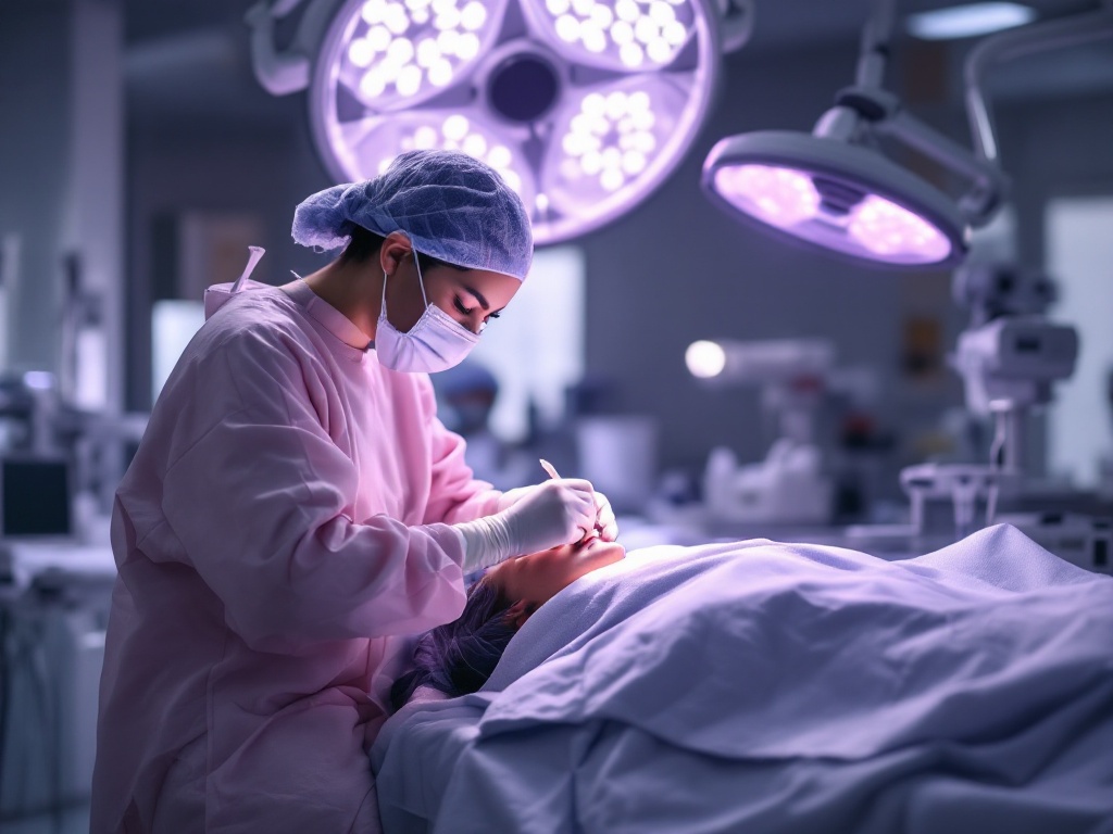
For many people living with a keloid, the instinct is understandable: just cut it off. The scar is raised, it itches or burns, it catches on clothing, and removal sounds like closure. But dermatologic surgeons have known for decades that excision by itself is one of the least reliable ways to treat a keloid. Published recurrence rates for surgery alone commonly fall in the range of 45 to 100 percent, and the regrown keloid is sometimes larger than the original. Understanding why requires looking at what a keloid actually is.
A keloid is a wound-healing process that never received the stop signal. Normal scars form when fibroblasts, the cells that produce collagen, respond to injury, lay down repair tissue, and then wind down. In keloid-prone skin, those fibroblasts stay switched on. They overproduce collagen, particularly type I, and they respond abnormally to signaling molecules such as transforming growth factor beta. The result is scar tissue that grows beyond the borders of the original wound and does not regress on its own.
This is the core problem with excision as a standalone treatment. Cutting out a keloid creates a fresh wound in skin that has already demonstrated, in the most direct way possible, that it heals abnormally. You are asking the same dysregulated fibroblasts to behave differently the second time, with no new instructions. Often they do not. They receive the same inflammatory signals, mount the same exaggerated response, and rebuild the keloid along the new incision line.
What adjuvant therapy actually does. The logic of combination treatment is to pair surgical removal, which debulks the lesion, with a second intervention that interrupts the biological loop driving regrowth. Several approaches have meaningful evidence behind them.
Intralesional corticosteroids, most commonly triamcinolone acetonide, are the workhorse. Injected into the wound edges after excision and repeated at intervals of roughly three to six weeks, steroids suppress inflammation, reduce fibroblast proliferation, and increase collagenase activity, which breaks down excess collagen. When excision is followed by a proper steroid injection series, reported recurrence rates typically drop below 50 percent, and in some series considerably lower. The tradeoffs are real: skin thinning, lightening of pigment, and visible small blood vessels can occur, particularly with repeated high doses.
Superficial radiation therapy delivered within the first 24 to 72 hours after excision has some of the strongest data of any adjuvant. Low-dose radiation damages the rapidly dividing fibroblasts that would otherwise repopulate the wound, essentially removing the workforce before construction restarts. Multiple studies report recurrence rates in the range of 10 to 30 percent when protocols are followed. The main hesitation is theoretical long-term cancer risk, which appears very low at the doses used but leads many clinicians to reserve radiation for earlobes, chest, and other sites away from radiosensitive tissue, and to avoid it in younger patients when alternatives exist.
5-fluorouracil, a chemotherapy agent used at low doses via injection, inhibits fibroblast proliferation through a different pathway than steroids. It is often mixed with triamcinolone, a combination that in several trials outperformed steroid alone and produced less skin atrophy. It is an off-label use and injections can be painful, but it has become a common second-line or combination tool.
Pressure therapy and silicone occupy the maintenance tier. Pressure earrings after earlobe keloid excision are a well-established example: sustained mechanical pressure appears to reduce blood flow and mechanotransduction signals in the healing tissue. Silicone sheeting or gel, worn for months, hydrates the scar surface and may modulate fibroblast behavior, though its effect size is modest. Neither is powerful alone against an established keloid, but both are low-risk additions after surgery.
What this means practically. If a clinician proposes removing your keloid, the most important question is not about the excision technique. It is: what is the plan for the weeks and months afterward? A credible answer names a specific adjuvant, a schedule, and a follow-up window, because keloid recurrence usually declares itself within the first year, and early regrowth is easier to intercept than a fully reformed lesion.
It also means managing expectations honestly. Even the best combination protocols do not guarantee zero recurrence. Keloid biology varies by anatomic site, skin type, genetics, and the tension across the wound. Chest, shoulders, and jawline are notoriously stubborn. Earlobes respond best.
The encouraging part is that the field has largely moved past the era of excision alone. The modern standard treats surgery as step one of a sequence, not a cure in itself. For patients, that reframing is the single most useful piece of knowledge to carry into a consultation: a keloid is not just extra tissue to remove, it is a process to interrupt.
Related reading: Why keloids come back, and how recurrence is prevented.