
Explainer · July 21, 2026 · 7 min · By Ifeoma Stanfield
How keloids are diagnosed, and when a biopsy is actually needed
Most keloids are diagnosed by eye and by history. Knowing the few situations that call for a biopsy, and why clinicians hesitate to cut, is the useful part.
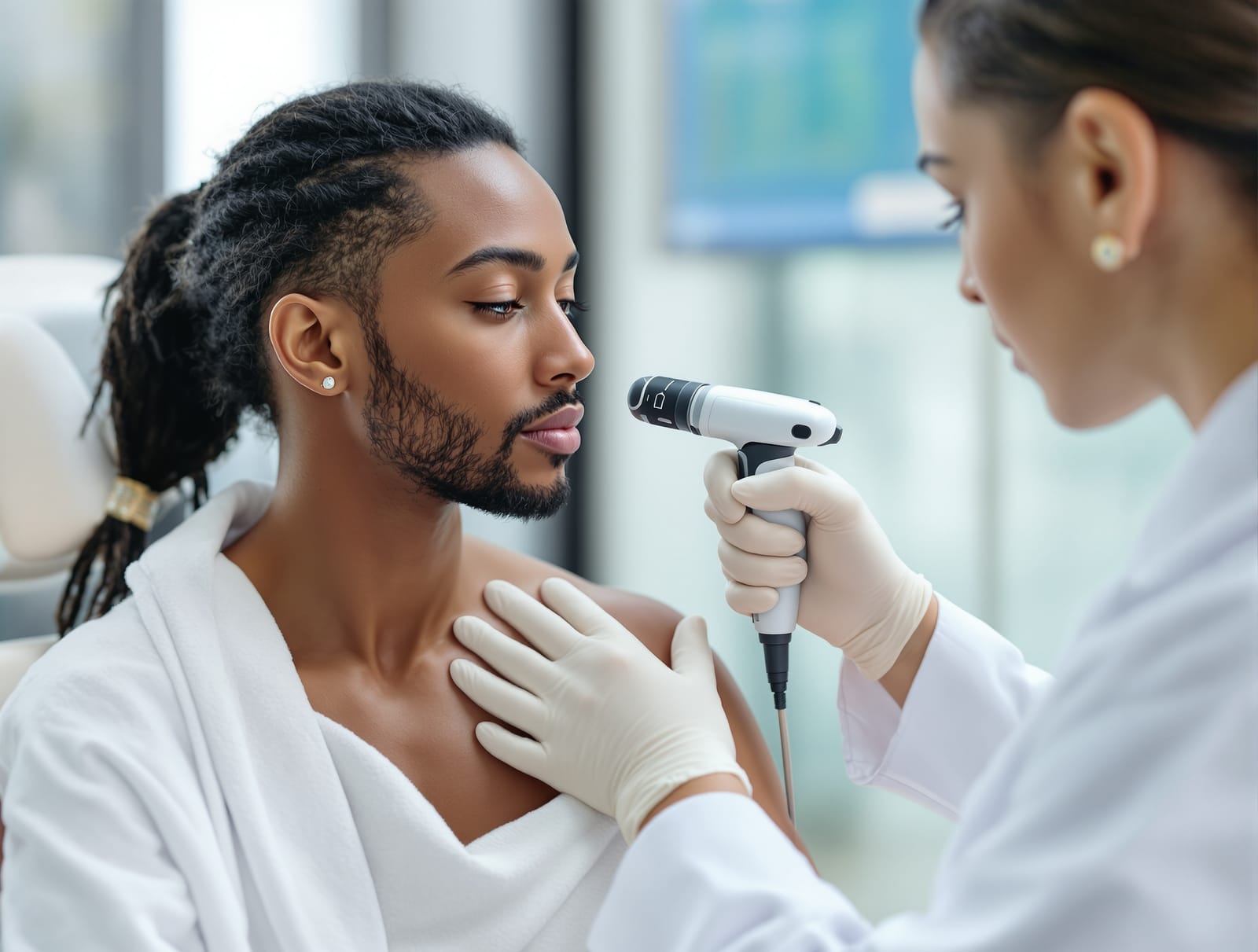
Patients often arrive at a dermatology appointment expecting a test. Blood work, a scan, a sample sent to a lab, something that returns a verdict. For keloids, that is usually not how it goes. The great majority of keloids are diagnosed clinically, meaning the dermatologist looks at the scar, presses on it, and asks a handful of pointed questions. That can feel underwhelming, but it reflects how distinctive a keloid is once you know what to look for, and it reflects a real reluctance to put a scalpel into skin that has already proven it scars excessively.
What the clinician is actually looking at. A keloid has a recognizable signature. It is firm and rubbery rather than soft, often smooth and shiny on the surface, and it is usually raised well above the surrounding skin. Color runs from pink or red in an active lesion to darker brown or purple, depending on skin tone and age of the scar. The defining feature, and the one the whole diagnosis rests on, is that a keloid extends beyond the boundaries of the original wound into skin that was never injured. That single detail is what separates a keloid from an ordinary or hypertrophic scar, and it is the first thing a dermatologist checks (StatPearls keloid overview, NIH).
The history matters as much as the exam. A good keloid consultation spends real time on questions rather than the lesion itself. When did it start, and what caused it? A keloid follows an injury, even a small one: a piercing, a vaccination, a surgical incision, a burn, an ingrown hair, or an inflamed acne lesion. Did it keep growing after the wound healed, and is it still growing now? Has it ever gone down on its own? Does it itch or hurt? Have you scarred like this before, and does anyone in your family? Those answers, combined with the site of the lesion and your risk profile, usually settle the diagnosis without any test at all (DermNet, keloid and hypertrophic scar).
Keloid or hypertrophic scar: the distinction that changes the plan. These two are confused constantly, including in clinical settings, and the difference is not academic. A hypertrophic scar stays inside the footprint of the original wound and often flattens and fades over one to two years without much intervention. A keloid spreads past the wound margin, does not reliably regress, and returns readily after simple removal. Getting this right determines whether you are told to wait and watch or enrolled in an active treatment course such as a series of intralesional steroid injections. Timing helps too: hypertrophic scars typically appear within weeks of an injury, while a keloid may take months to declare itself and then continue enlarging long afterward.
Why a biopsy is not routine. Here is the part that surprises people. Cutting into a keloid to sample it creates a fresh wound in exactly the tissue most likely to overgrow, and that biopsy site can itself keloid, sometimes leaving a bigger problem than the question the biopsy was meant to answer. Dermatologists therefore treat a biopsy as a decision with a real cost rather than a free source of information. When one is genuinely necessary, an experienced clinician plans for it: taking a small sample, closing under low tension, and pairing it with a prevention plan such as silicone, pressure, or an injection into the healing site.
When a biopsy is warranted. There is a short list of situations where the tissue answer is worth the wound. The clearest is a lesion that does not fit the story. If a firm growing nodule appeared with no preceding injury, in a site and a person with no keloid history, the working assumption that it is a keloid weakens. Rapid or asymmetric growth, ulceration or a surface that breaks down and does not heal, bleeding, drainage, a change in texture or firmness in one part of an old stable scar, or a lesion that keeps expanding despite adequate treatment all raise the same question. So does a keloid that has been present and unchanged for many years and then suddenly starts behaving differently.
What clinicians are ruling out. Several conditions can imitate a raised, firm scar. Dermatofibrosarcoma protuberans is a rare, slow-growing soft tissue tumor that begins as a firm plaque or nodule and is repeatedly mistaken for a keloid or a benign scar, which is a well-recognized cause of delayed diagnosis (DermNet, dermatofibrosarcoma protuberans). Other mimics include dermatofibroma, hypertrophic scar, foreign body granuloma from retained material in an old wound, and in long-standing chronically ulcerated scars, squamous cell carcinoma. None of these are common, and none of them should make you assume the worst about a typical keloid, but they are the reason a scar behaving strangely earns a closer look instead of another round of injections.
Other tools, and their limits. Imaging is not part of standard keloid diagnosis. Ultrasound is sometimes used in research settings or by specialist centers to measure scar thickness and track response over a treatment course, and MRI may be ordered if a deeper mass is suspected rather than a surface scar. Photography, by contrast, is genuinely useful and underused: dated photographs taken at each visit turn slow month over month change into something you can actually see, which matters enormously in a condition where progress is measured in softening and flattening over many months.
What to bring to the appointment. You can make the clinical diagnosis faster and more accurate with very little effort. Bring the date and cause of the original injury if you know it, any photographs showing the scar at earlier points, a list of what you have already tried and for how long, your personal and family history of unusual scarring, and a note of whether the lesion itches, hurts, or has changed recently. If you already know you are in a higher-risk group, say so early, because it shapes both the diagnosis and the prevention plan that follows.
The takeaway. A keloid is almost always diagnosed by examination and history rather than by a test, and the single most important finding is a scar that has grown beyond the wound that caused it. A biopsy is not a routine step and carries the specific risk of provoking a new keloid at the sampling site, so it is reserved for lesions that do not fit the pattern: growths with no preceding injury, rapid or asymmetric enlargement, ulceration or bleeding, or a stable old scar that suddenly changes. If your lesion looks and behaves like a keloid, expect a treatment plan rather than a lab result. If it does not, ask directly what else is on the list and whether sampling it is worth the wound.
Related reading: What is a keloid, and how is it different from a scar? and Who gets keloids, and why.