
Explainer · August 7, 2026 · 4 min · By Ifeoma Stanfield
Steroid or 5-FU? What the Evidence Says About the Two Most Common Keloid Injections
Intralesional triamcinolone has been the default keloid injection for decades, but 5-fluorouracil is increasingly used alongside or instead of it. Here is how each drug works, what the trials show, and why many clinicians now combine them.
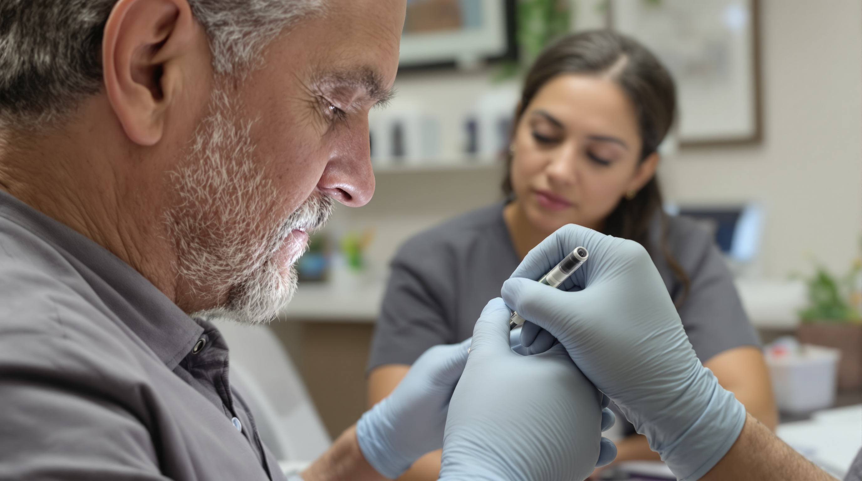
If you have been evaluated for a keloid, there is a good chance someone offered you an injection. For most patients, that injection is triamcinolone acetonide, a corticosteroid delivered directly into the scar. Increasingly, though, dermatologists are reaching for a second drug, 5-fluorouracil (5-FU), either on its own or mixed in the same syringe. Understanding what each agent actually does inside a keloid helps explain why the field is shifting, and why neither is a magic fix.
A keloid is not simply a thick scar. It is a benign but persistently active growth of fibroblasts, the cells that manufacture collagen. In a keloid, those fibroblasts behave as if the wound never closed. They overproduce collagen, particularly type I, and they resist the normal signals that tell scar tissue to stop growing and remodel. Any effective injectable has to interrupt that loop, either by calming the inflammatory signaling that drives fibroblasts or by slowing the fibroblasts themselves.
How triamcinolone works. Corticosteroids act on several fronts at once. They suppress inflammatory mediators, reduce the activity of transforming growth factor beta, which is a key driver of collagen production, and increase collagenase activity, which breaks down existing collagen. Clinically, that translates into softening and flattening of the keloid over a series of treatments, typically spaced three to six weeks apart. Response rates in published series generally fall in the range of 50 to 90 percent for meaningful flattening, though recurrence after stopping treatment is common, often reported between 10 and 50 percent depending on follow-up length and keloid location.
The trade-offs are well documented. Steroid injections can cause skin atrophy, visible thinning of the surrounding tissue, along with hypopigmentation, which is particularly noticeable in darker skin tones, and telangiectasias, the fine surface blood vessels that appear when the dermis thins. These effects are dose related, which is why clinicians titrate concentration, commonly between 10 and 40 milligrams per milliliter, and limit total volume per session.
How 5-FU works. 5-fluorouracil is a chemotherapy agent, and that word understandably alarms patients, but the mechanism at intralesional doses is targeted rather than systemic. 5-FU interferes with DNA synthesis in rapidly dividing cells. In a keloid, the most rapidly dividing cells are the overactive fibroblasts. The drug slows their proliferation and reduces collagen type I production, essentially turning down the factory rather than demolishing the product. The total dose used in a keloid session is a small fraction of what oncologists use, and systemic side effects are rare in published dermatologic series.
What 5-FU does not do is thin the skin. Because it works on cell division rather than on the structural matrix directly, atrophy and hypopigmentation are far less common than with steroids. Its typical side effects are local: injection pain, temporary darkening at the site, and occasionally small ulcerations that heal on their own. Patients who are pregnant or have significant anemia or bone marrow suppression are generally not candidates.
What head-to-head data show. Randomized comparisons are modest in size but reasonably consistent. Trials comparing triamcinolone alone, 5-FU alone, and the two combined tend to find that the combination outperforms either drug by itself, both in the degree of flattening and in patient-reported symptoms like itch and tenderness. A frequently cited approach mixes a small amount of triamcinolone into a 5-FU syringe, often around a 1 to 9 ratio, so the steroid dose per session drops substantially. That lower steroid exposure appears to reduce atrophy and pigment changes while the two mechanisms, anti-inflammatory plus antiproliferative, reinforce each other. Several systematic reviews have reached the same general conclusion, while noting that study protocols vary widely in concentration, interval, and number of sessions, which makes precise effect sizes hard to pin down.
What this means practically. Neither injection cures a keloid in the sense of removing it. Both aim to flatten, soften, and quiet the scar, and both require multiple sessions, commonly three to eight, before judging success. Recurrence remains possible with any monotherapy, which is why injections are often one component of a plan that may include silicone sheeting, pressure therapy for ear keloids, or surgery with adjuvant treatment for large lesions.
A few honest caveats. Response varies by keloid age, size, and location; earlobe keloids tend to respond better than chest or shoulder keloids. Injection technique matters, since drug deposited under the keloid rather than into it does little. And pain is real for both agents, though many clinicians use topical anesthetic or mix lidocaine into the syringe.
The bottom line: triamcinolone remains a reasonable first-line injection, but if you have had atrophy or pigment loss from steroids, or your keloid has plateaued, asking about 5-FU or a combined protocol is a well-supported question, not a fringe one. The mechanisms are complementary, the evidence base, while imperfect, points the same direction, and the conversation is worth having before concluding that injections have failed you.