
Explainer · August 6, 2026 · 5 min · By Ifeoma Stanfield
Why Steroid Injections Sometimes Stop Working on Keloids, and What Clinicians Do Next
Intralesional triamcinolone remains the first-line keloid treatment worldwide, yet a meaningful share of scars plateau or rebound. Here is what is actually happening in the tissue, and how treatment plans adapt when the needle alone is not enough.
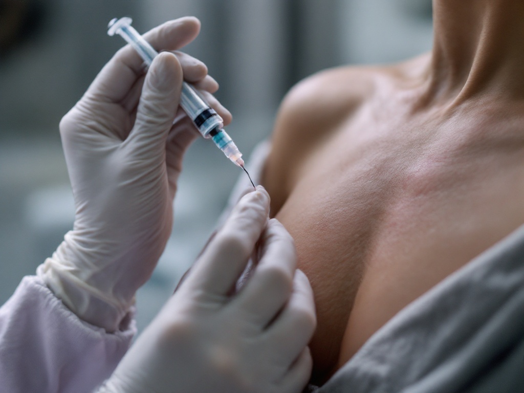
Ask almost any dermatologist how they start treating a keloid and the answer is the same: intralesional corticosteroid injections, usually triamcinolone acetonide. It is inexpensive, widely available, and supported by decades of clinical use. But patients often discover a frustrating pattern. The first two or three sessions soften and flatten the scar noticeably, then progress slows, stalls, or reverses after treatment stops. Understanding why requires looking at what the drug does and does not do inside a keloid.
How triamcinolone actually works. A keloid is not simply excess scar tissue. It is an active, chronically inflamed lesion in which fibroblasts overproduce collagen, particularly type I, and underproduce the enzymes that break collagen down. Triamcinolone attacks this on several fronts. It suppresses inflammatory signaling, reduces fibroblast proliferation, decreases collagen synthesis, and increases collagenase activity by blocking the natural inhibitors of that enzyme. The visible result is a scar that becomes softer, flatter, less itchy, and less painful over a series of injections spaced roughly three to six weeks apart.
Why the effect plateaus. Several mechanisms explain diminishing returns. First, older keloids contain dense, poorly vascularized collagen bundles at their core. Steroid suspension injected into that tissue distributes unevenly, and the deepest fibrotic zones may receive little drug at all. This is one reason clinicians often see the edges of a keloid respond while a firm central nodule persists. Second, keloid fibroblasts are not a uniform population. Research on cultured keloid cells suggests subsets vary in their sensitivity to glucocorticoids, so repeated treatment may preferentially suppress responsive cells while resistant ones continue producing matrix. Third, the underlying drivers, including skin tension, genetic predisposition, and in some cases ongoing irritation from hair follicles or piercings, are untouched by the injection. The drug quiets the output of the process without removing its cause.
Rebound after stopping. Keloids are defined by their tendency to recur, and steroid monotherapy has a recurrence rate commonly cited in the range of roughly 30 to 50 percent within a few years. When injections stop, the inflammatory and fibrotic signaling that was suppressed can resume, particularly in high-tension areas such as the chest, shoulders, and upper back.
What clinicians do when the needle stalls. The modern approach is combination therapy, and the evidence increasingly supports it over any single modality.
The most common pairing is triamcinolone with 5-fluorouracil (5-FU), a chemotherapy agent that blocks fibroblast proliferation through a different mechanism, interfering with DNA synthesis. Mixed in the same syringe, typically at a low steroid concentration, the combination has shown better flattening and fewer steroid side effects such as skin thinning, visible blood vessels, and pigment lightening in multiple comparative trials.
Cryotherapy before injection is another practical adjunct. A brief freeze softens the scar and creates edema, which allows the steroid to distribute more evenly through previously impenetrable tissue. Intralesional cryotherapy, where a probe freezes the keloid from the inside out, targets the core directly and tends to spare surface pigment better than spray freezing, an important consideration for patients with darker skin tones, who are both more prone to keloids and more prone to pigment changes from treatment.
For bulky or long-standing keloids, surgical excision followed immediately by adjuvant therapy is often the more realistic path. Excision alone is a known trap, with recurrence rates historically reported between roughly 45 and 100 percent, and the regrown keloid can exceed the original. But excision followed by superficial radiotherapy or brachytherapy within the first day or two, or by a structured course of postoperative steroid injections, brings recurrence down substantially in published series. Radiation works by impairing the proliferation of the fibroblasts that would otherwise rebuild the keloid during early wound healing.
Laser-assisted approaches are also gaining ground. Fractional ablative lasers create microscopic channels that improve drug delivery into dense scar tissue, and pulsed dye lasers target the blood vessels that feed active keloids, reducing redness and possibly the metabolic supply of the lesion.
What patients should take from this. A stalled response to injections is common and does not mean the keloid is untreatable. It usually means the treatment plan needs another mechanism added, not more of the same. Reasonable questions to raise with a clinician include whether 5-FU can be added, whether cryotherapy or laser pretreatment might improve drug penetration, and, for large or recurrent lesions, whether excision with immediate adjuvant therapy is appropriate. It is also worth confirming the basics: injections placed into the scar itself rather than beneath it, adequate but not excessive concentration, and consistent intervals.
Keloids are a chronic condition of wound healing, not a one-time defect. The most durable results come from layered strategies, realistic timelines measured in months, and follow-up long enough to catch early recurrence when it is still easy to treat.
Related reading: Steroid injections: the first-line keloid treatment.